e anatomy imaios|imioas enatomy : iloilo e-Anatomy is a comprehensive online resource for human anatomy and medical imaging. It offers anatomical diagrams, CT, MRI, radiographs, endoscopy, . web6 de jun. de 2022 · Para ajudar você nessa tarefa, o GUIA DO ESTUDANTE reuniu as provas e os gabaritos de todas as edições do vestibulinho das Etecs – lembrando que do segundo semestre de 2020 ao primeiro de .
0 · imioas enatomy
1 · imaos vet anatomy
2 · imaios free brain anatomy
3 · imaios e anatomy free
4 · imaios brain anatomy
5 · e anatomy sign in
6 · e anatomy free download
7 · brain images anatomy
webGier - Gestão Inteligente da Educação Responsável. seja bem-vindo. Proceda seu acesso por usuário: IDENTIFICAÇÃO POR USUÁRIO.
e anatomy imaios*******e-Anatomy is a comprehensive online resource for human anatomy and medical imaging. It offers anatomical diagrams, CT, MRI, radiographs, endoscopy, .Orbit Mri Mri Premium - e-Anatomy, the Anatomy of Imaging - IMAIOSAnnotated dental-CBCT - e-Anatomy, the Anatomy of Imaging - IMAIOSIntroduction. This anatomical module of e-Anatomy is dedicated to the anatomy of .CT Arthrography - e-Anatomy, the Anatomy of Imaging - IMAIOSIMAIOS delivers high-quality anatomy and imaging content for daily practice and training of health professionals, guides accurate diagnosis and reporting through detailed .imioas enatomy We created a brain atlas that is an interactive tool for studying the conventional anatomy of the normal brain based on a magnetic resonance imaging exam of the axial brain. Anatomical .
IMAIOS e-Anatomy is an atlas of human anatomy for physicians, radiologists, medical students and radiology technicians. .IMAIOS e-Anatomy is an atlas of human anatomy for physicians, radiologists, medical students and radiology technicians. Get a sneak peek at more than 26 000 medical and .
To access to all e-Anatomy modules you must log in using an account that has an active e-Anatomy subscription. If you wish to subscribe, please follow the following steps: 1. Log .e-Anatomy is an interactive human anatomy database designed for physicians, radiologists, and medical students. It includes over 40 different modules that feature thousands of .
How can I access all the modules and features of e-Anatomy? To access to all e-Anatomy modules you must log in using an account that has an active e-Anatomy subscription. If .Our user guide is available for download here: User Guide. This guide is also available in other languages: Deutsch: Benutzerhandbuch. Español: Manual de instrucciones. . This page presents a comprehensive series of labeled axial, sagittal and coronal images from a normal human brain magnetic resonance imaging exam. This MRI brain cross-sectional anatomy tool .
This cross-sectional human anatomy atlas of the lower limb is an interactive tool based on MRI axial images of the human leg. Anatomical structures of the lower limb (hip, thigh, knee, leg, anke and .The foot serves as the terminal part of the limb responsible for bearing weight and enabling locomotion. Its skeletal structure comprises three main components: the tarsus, metatarsus, and phalanges.Tarsus:The tarsus .

MRI of the shoulder : muscles of the rotator cuff labeled on a sagittal MR slice. An MRI of the shoulder of a healthy subject was performed in the 3 planes of space (coronal, axial, sagittal) commonly .

MRI of the shoulder : muscles of the rotator cuff labeled on a sagittal MR slice. An MRI of the shoulder of a healthy subject was performed in the 3 planes of space (coronal, axial, sagittal) commonly . Anatomy of the hip (cross-sectional imaging on 3T MR and 3D medical pictures) Images provided by Sorin Ghiea & Emi Preda - MD. This radioanatomy atlas is about the articulation and the hip area on MRI. On these 252 3T MRI images over 340 anatomical structures were labeled. At the end of this module, there are 3D .The abdomen is a body region which is cylindrical in shape and is situated between the thorax (above) and the pelvis (below). The abdominal cavity extends from the diaphragm above, to the pelvic inlet or pelvic brim below. In fact, the abdominal and pelvic cavities are continuous entities, forming one large abdominopelvic peritoneal cavity. Outside, the . Anatomy of the head on a cranial CT Scan: brain, bones of cranium, sinuses of the face. Coronal Brain CT. Vasculary territories. Dural venous sinuses, Veins, Arteries. Bones of cranium Axial CT. Paranasal sinuses - CT. Cranial base , CT: Foramina, Nasal cavity, Paranasal sinuses. Bones of cranium : Anatomy , CT. ANATOMICAL .
Lymph nodes of the face, neck, thorax, abdomen and pelvis - hepatic segmentation - entire body scan in oncology. This human anatomy module is about the lymph nodes, ganglionic areas and organs involved in oncological disease spread assessments. It was created from a scanner (computed tomography) with iodine .
The ankle, or the talocrural region, is the region where the foot and the leg meet.The ankle includes three joints: the ankle joint proper or talocrural joint, the subtalar joint, and the inferior tibiofibular joint.[The movements produced at this joint are dorsiflexion and plantarflexion of the foot. In common usage, the term ankle refers exclusively to the .Gallery. The lungs are the essential organs of respiration; they are two in number, placed one on either side within the thorax, and separated from each other by the heart and other contents of the mediastinum. The substance of the lung is of a light, porous, spongy texture; it floats in water, and crepitates when handled, owing to the presence .
IMAIOS and selected third parties, use cookies or similar technologies, in particular for audience measurement. Cookies allow us to analyze and store information such as the characteristics of your device as well as certain personal data (e.g., IP addresses, navigation, usage or geolocation data, unique identifiers).
1. Submental triangle, 2. Submandibular triangle –housing the submandibular gland, facial vein and artery, 3. Carotid triangle –housing the carotid artery and its branches, and the internal jugular vein and vagus nerve, and. 4. Muscular triangle –housing the infrahyoid strap muscles of neck. The posterior triangle is further subdivided . ENT anatomy: MRI of the face and neck - interactive atlas of human anatomy using cross-sectional imaging. We attempted to synthesize the anatomy of the face and neck in this anatomy module. We used MRI images T2-weighted with axial, sagittal and coronal planes. 512 anatomical structures were dynamically labeled, and some .
Anatomy of the face and neck (CT) - interactive atlas of human anatomy using cross-sectional imaging. This head and neck anatomy atlas is an educational tool for studying the normal anatomy of the face based on a contrast enhanced multidetector computed tomography imaging (axial and coronal planes). Interactive labeled images .e anatomy imaiosIMAIOS delivers high-quality anatomy and imaging content for daily practice and training of health professionals, guides accurate diagnosis and reporting through detailed anatomical views and multiple modalities to empower understanding and confidence at every career stage. Available on the web and mobile.e anatomy imaios imioas enatomy e-Anatomy delivers a high quality anatomy and imaging content atlas. It is the most complete reference of human anatomy available on the , iOS and Android devices. Pinpoints Detailed Views Across Anatomical Regions & Modalities (CT, MRI, Radiographs), Anatomic diagrams and nuclear images.IMAIOS delivers high-quality anatomy and imaging content for daily practice and training of health professionals, guides accurate diagnosis and reporting through detailed anatomical views and multiple modalities to empower understanding and confidence at .
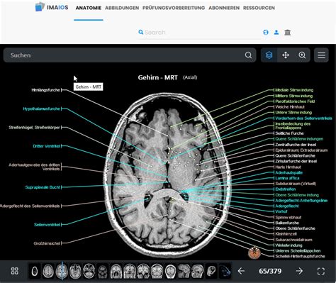
We created a brain atlas that is an interactive tool for studying the conventional anatomy of the normal brain based on a magnetic resonance imaging exam of the axial brain. Anatomical structures and specific areas are .
IMAIOS e-Anatomy is an atlas of human anatomy for physicians, radiologists, medical students and radiology technicians. Get a sneak peek at more than 26 000 medical and anatomical images.IMAIOS e-Anatomy is an atlas of human anatomy for physicians, radiologists, medical students and radiology technicians. Get a sneak peek at more than 26 000 medical and anatomical images for free before subscribing to our detailed atlas of human anatomy.
To access to all e-Anatomy modules you must log in using an account that has an active e-Anatomy subscription. If you wish to subscribe, please follow the following steps: 1. Log in on the IMAIOS website. 2. Click on “Subscribe” on the menu at the top of the page. 3.e-Anatomy is an interactive human anatomy database designed for physicians, radiologists, and medical students. It includes over 40 different modules that feature thousands of images of the human body. Available online to authorized COM/HMC users only. Access e-Anatomy.How can I access all the modules and features of e-Anatomy? To access to all e-Anatomy modules you must log in using an account that has an active e-Anatomy subscription. If you wish to subscribe, please follow the following steps: 1. Log in on the IMAIOS w. Continue reading; What are the free e-Anatomy modules?
14 de out. de 2023 · Máscara de payaso del video 'Quiero agua'. Foto: YouTube. El video supuestamente es de origen mexicano, aunque aún no hay muchos detalles detrás de .
e anatomy imaios|imioas enatomy