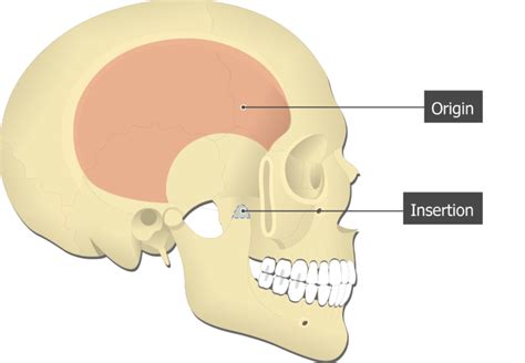
temporalis origin and insertion|temporalis muscle diagram : Clark Origin and insertion. Temporalis muscle. Musculus temporalis. 1/5. Synonyms: Temporal muscle. The temporalis muscle is a broad muscle that occupies most of the temporal fossa. Its origin point spans the entire surface of the fossa below the . Resultado da Marcinha Caminhoneira. 40,722 likes · 817 talking about this. Entrepreneur.
0 · where is the temporal tendon
1 · temporalis muscle picture
2 · temporalis muscle diagram
3 · temporalis function and location
4 · temporalis fascia attaches to
5 · temporal fossa muscle diagram
6 · muscle that inserts in jaw
7 · latissimus dorsi origin and insertion
8 · More
Resultado da Well, if you're ready to embark on this powerful journey, let Movida Escorts be your guide as we unveil the secrets to becoming a successful London .
temporalis origin and insertion*******Origin and insertion. Temporalis muscle. Musculus temporalis. 1/5. Synonyms: Temporal muscle. The temporalis muscle is a broad muscle that occupies most of the temporal fossa. Its origin point spans the entire surface of the fossa below the .
In humans, the temporalis muscle arises from the temporal fossa and the deep part of temporal fascia. This is a very broad area of attachment. It passes medial to the zygomatic arch. It forms a tendon which inserts onto the coronoid process of the mandible, with its insertion extending into the retromolar fossa posterior to the most distal mandibular molar. In other mammals, the muscle usually spans the dorsal part of the skull all the way up to the medial line. There, it may be attac. Learn about the temporalis muscle, a broad and thin muscle of mastication located at the side of the head. Find out its origin, insertion, actions and innervation, and test yourself with an interactive quiz. The temporalis muscle is a broad, fan-shaped muscle that fills much of the temporal fossa. It arises from the inferior temporal line on the bony floor of the temporal .
Learn about the temporalis muscle, a broad and thin muscle that helps in mastication and jaw movements. Find out its location, origin, insertion, function, innervation, and blood supply in this lesson. The temporalis is a muscle of mastication (chewing). It is located on the lateral aspect of the skull. Attachments: Originates from the temporal fossa of the skull .
Origin: Temporal fossa and fascia. Insertion: Coronoid process and ramus of mandible. Action: Elevates and retracts mandible. Innervation: Anterior and posterior .
The temporalis muscle is one of the four primary muscles of mastication (chewing of food). It is a fan-shaped muscle with anterior fibres that have a vertical orientation, mid fibres .Origin and insertion of temporalis by Anatomy.app . Insertion. The temporalis forms a tendon that inserts onto the coronoid process of the mandible. Action. Upon activation, the temporalis muscle elevates the .

Origin: Greater part of the temporal fossa, between the lower temporal line and the infratemporal crest (frontal, sphenoid and parietal bone) and on the medial part of the . Medial pterygoid is a thick quadrilateral muscle that connects the mandible with maxilla, sphenoid and palatine bones. It belongs to the group of masticatory muscles, along with the lateral pterygoid, masseter .
The Temporalis Muscle. Origin: Temporal fossa and temporal fascia. The fibers blend together to pass between the zygomatic arch and the side of the skull. Insertion: The upper, anterior border and inner surface of the .Origin: Parietal, temporal, frontal and occipital bones.Insertion: Medial surface of the condyle of the mandible just ventral to its articular surface.Action: Raises the mandible.Nerve: Trigeminus. Introduction. The temporalis muscle (TM) is composed of a single layer within the temporal line of the parietal bone and attaches to the coronoid process [1-4].In major textbooks and atlases, it is usually illustrated simply as originating at a wide attachment site, converging to the form of a tendon, and inserting onto a narrow site of .The masseter and temporalis muscles are palpated on both sides. The masseter is palpated under the zygomatic process on the lateral cheek above the angle of the jaw. The temporalis muscle is palpated over the temple at the hairline, anterior to the ear and superior to the zygomatic bone. 0: Patient cannot completely close the mouth.
Summary. origin: temporal fossa between the infratemporal crest and inferior temporal line on the parietal bone; deep surface of the temporalis fascia. insertion: coronoid process and ramus of mandible. innervation: deep temporal nerves, branches off the anterior division of the mandibular division of the trigeminal nerve. action: elevate .
Muscles of mastication (Masticatory muscles) The muscles of mastication are a group of muscles that consist of the temporalis, masseter, medial pterygoid and lateral pterygoid muscles.The temporalis muscle is situated in the temporal fossa, the masseter muscle in the cheek area, while the medial and lateral pterygoids lie in the .M. Temporalis origin From fossa temporalis and its borders: linea temporalis, external Sagittarius crest, nuchal crest and dorsal edge of zygomatic arch M. Temporalis insertionTerms in this set (3) orgin of temporalis. temporal bone. insertion of temporalis. coronoid process and lateral surface of mandible. temporalis action. elevates mandible. Study with Quizlet and memorize flashcards containing terms like orgin of temporalis, insertion of temporalis, temporalis action and more.
Origin of Temporalis Muscle. The temporalis muscle is a wide muscle that occupies most of the temporal fossa. Its origin point traverses the whole surface of the fossa below the temporal line. Additionally, some fibers arise from the temporal fascia as well. Insertion. The temporalis muscles are divided into the anterior and posterior .
TEMPORALIS. ORIGIN Temporal fossa between inferior temporal line and infratemporal crest: INSERTION Medial and anterior aspect of coronoid process of mandible: ACTION Elevates mandible and posterior fibers retract: NERVE Deep temporal branches from anterior division of mandibular nerve (V) .
temporalis origin and insertion Lateral pterygoid is located deep to the temporalis and masseter muscles, spanning between the sphenoid bone and temporomandibular joint. Its muscle belly is separated by a small horizontal fissure into two heads; superior (upper) and inferior (lower). The superior head is formed by the most superomedial fibers of the muscle. There are four muscles: Masseter. Temporalis. Medial pterygoid. Lateral pterygoid. The muscles of mastication develop from the first pharyngeal arch. They are therefore innervated by a branch of the trigeminal nerve (CN V), the mandibular nerve. In this article, we shall look at the anatomy of the muscles of mastication – their .

The origin, insertion, and action of the main muscles of mastication, as well as a brief description of the accessory muscles of mastication, are as follows. Temporalis Muscle The temporalis muscle is a fan-shaped muscle with anterior fibers that have a vertical orientation, mid fibers have an oblique orientation, and posterior fibers have a .
TEMPORALIS. ORIGIN Temporal fossa between inferior temporal line and infratemporal crest: INSERTION Medial and anterior aspect of coronoid process of mandible: ACTION Elevates mandible and posterior fibers retract: NERVE Deep temporal branches from anterior division of mandibular nerve (V) . Lateral pterygoid is located deep to the temporalis and masseter muscles, spanning between the sphenoid bone and temporomandibular joint. Its muscle belly is separated by a small .
There are four muscles: Masseter. Temporalis. Medial pterygoid. Lateral pterygoid. The muscles of mastication develop from the first pharyngeal arch. They are therefore innervated by a branch of the .temporalis origin and insertion temporalis muscle diagram The origin, insertion, and action of the main muscles of mastication, as well as a brief description of the accessory muscles of mastication, are as follows. Temporalis Muscle The temporalis muscle is a fan-shaped muscle with anterior fibers that have a vertical orientation, mid fibers have an oblique orientation, and posterior fibers have a .
Origin of Temporalis muscle. Temporal fossa, excluding the zygomatic bone, Temporal fascia. Insertion of Temporalis muscle. The fibers of the temporalis muscle converge and pass through the gap deep to the zygomatic arch. They are inserted into The margins and deep surface of the coronoid process; and the anterior border of .Elevation and retrusion of mandible. What is the unilateral movement of the Temporalis muscle? Lateral excursion to the ipsilateral side. Study with Quizlet and memorize flashcards containing terms like What is the temporalis muscle origin?, What is the Temporalis insertion?, What is the nerve innervation for the Temporalis muscle? and . In addition to the temporalis, the lateral pterygoid, and the medial pterygoid, the masseter represents one of four muscles of mastication or chewing. The masseter muscle represents the most .Match the muscle with its correct origin and insertion: Temporalis. Origin: temporal fossa Insertion: coronoid process of mandible. Identify the muscle indicated by "E." (back) Supraspinatus. Identify the muscle indicated by "F." (back) Infraspinatus. Origin. Insertion. Action. Innervation. Deep masseter. Zygomatic arch. Mandible. Raises mandible against the maxillae with great force. Trigeminal nerve (V), mandibular branch (V3) Temporalis. Temporal fossa and temporal fascia. Coronoid process and anterior ramus of the mandible. Elevates and retracts the mandible against .temporalis muscle diagram Temporalis Muscle : Origin, Insertion, Nerve supply, Clinical importance - Anatomymuscle of mastication. Temporalis. origin: temporal fossa. insertion: coronoid process and ramus of mandible. action: elevation (shortens as mouth closes) innervation: trigeminal. muscle of mastication. Medial pterygoid. Origin: lateral pterygoid plate of the sphenoid bone and tuberosity of the maxilla.Insertion. buccinator. pressing cheek inward, compressing air while blowing. maxilla bone, mandible bone. orbicularis oris. depressor anguli oris. opening the mouth, sliding the lower jaw right and left. mandible bone. corners of the mouth.
web10 de jan. de 2020 · Gelobel, Londrina: See 685 unbiased reviews of Gelobel, rated 4.5 of 5 on Tripadvisor and ranked #13 of 967 restaurants in Londrina.
temporalis origin and insertion|temporalis muscle diagram